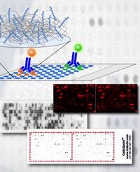
CelluSpots™ arrays made by Pepmic can be analyzed with standard chromogenic substrate reactions as used in Western blot protocols. Development is done in a small open tray. Spot intensities are recorded on a standard flatbed scanner. Detection by luminescence or radioactivity is also possible. Microarrays with smaller spots are available on request. These usually require analysis equipment and procedures as used for DNA microarrays.
-
standard format is a 26x76 mm glass slide covered with white adhesive foil
-
up to 384 peptide spots printed in duplicate on the foil side of the slide
-
spot-to-spot distance 1.2 mm
-
peptides are covalently bound to cellulose via C-terminus
-
N-terminus is usually blocked (acetylation)
-
arrays contain control peptides and location marks
Different CelluSpots™ array layouts are available and customized formats can be generated.
CelluSpots™ array, 2x384 spots:
CelluSpots™ array, 4x192 spots:
|