From Membranes to Mini- and Micro-Arrays
The SPOT synthesis technology is well known as a rapid and robust method to generate peptide libraries on membrane supports – particularly on cellulose sheets. Pepmic has many years of experience in the development of peptide synthesis, fully automated SPOT synthesis and parallel synthesis of peptides in a microtiter plate format.
Even though peptide libraries on SPOT membranes are popular in a wide range of screening methods there are three major drawbacks:
-
The re-usability of SPOT membranes is limited. For some assays they can be used only once.
-
The production of duplicate peptide SPOT arrays with identical quality is time-consuming and expensive as each membrane must be synthesized individually.
-
Membranes are large compared to micro arrays on glass slides and require large sample volumes.
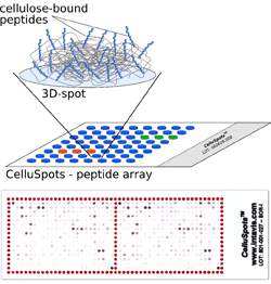
Pepmic now offers CelluSpots™ as a new format to use the advantages of the method but avoid the limitations of SPOT membranes. CelluSpots™ are arrays of peptide-cellulose conjugates spotted on planar surfaces such as glass slides. Peptides are synthesized on a modified cellulose support which can be dissolved after synthesis. The solutions of individual peptides covalently linked to macro molecular cellulose are then spotted onto a surface of choice. After evaporation of the solvent a three-dimensional layer is formed which is not dissolved in aqueous reagents used for standard assays. The three-dimensional structure holds up to 1000 times more peptides per area as compared to conventional monolayer deposition. This shifts the binding equilibrium in a favorable direction for low affinity protein-protein interactions.
Advantages of CelluSpots™ Arrays

-
numerous identical copies of the same quality can be prepared
-
smaller sample volumes are needed
-
high peptide density allows detection of low affinity interactions
-
chemical properties are comparable to those of the original SPOT membranes
-
detection by chemiluminescence, autoradiography or enzymatic color development
-
compatible with standard equipment for micro-arrays, e.g. hybridization chambers and scanners
-
low non-specific protein binding of cellulose
|